ACL TEAR
- Home
- ACL TEAR
What Is the Anatomy of the ACL Tear?
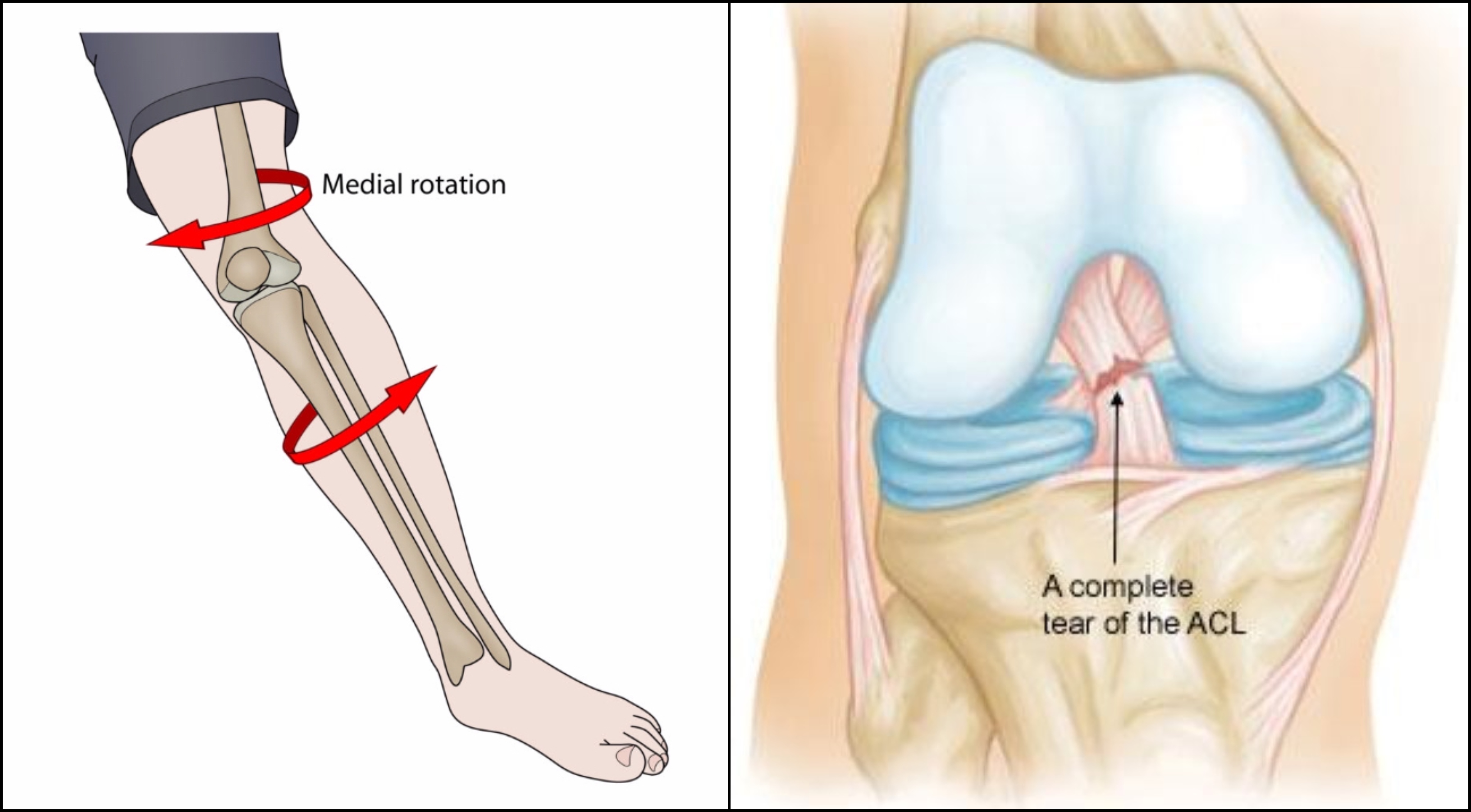
The ACL (anterior cruciate ligament) is the damaged knee ligament or element. The ACL tear connects the condyle to the tibia which is in front of the anterior tibial bone. Some of its tissues also combine into the middle meniscus.So, There are usually two bunches of tissues that give up the ACL and support it to maintain sustain the knee in bending, rotation, extension, and movement.Damaged ligaments or elements are also called strains. Therefore, They can group based on their hardness and different type of pain. As a result, ACL tear injury can classify into 3 grades.1. Grade 1: Ligament tissues are tightened or stretched although not ripped(Torn).2. Grade 2: Strains have some tissues are torn while the ligament works remain functionally sound.3. Grade 3: Injury occurs while the ligament is completely ripped and having pain and difficulty to do a normal activity or movement of the knee.
What Are Causes and Risk Factors of an ACL Tear?
When a person changing knee direction immediately and getting a sudden stop to run or arriving from a jump to height, With the legs, then the knee having over straightens and turns at the same time, accentuating the ACL and creating it to strain and tear. ACL tear injury happens when playing High-risk sports like soccer, basketball, cricket, and skiing.Women have a higher risk of an ACL injury or damaged than men. Possible analyses reason for having ACL tear injury in a woman may involve variances in anatomy, and movement experience. Some other reason for raised ACL tear injury in females is Genetic variances in how tissues arrangement. Moreover, women have an extended abdomen than men to provide childbirth, and this can increase the angle of the femur where they meet the tibia at the knee joint. An extended angle increases the intensity of the ACL tear in a knee ligament.
What Are the Symptoms of Torn ACL?
The sufferer normally can feel or hear a loud crunching and click sound in the ligament.
Observers seldom say that they can hear easily. The injury is almost instant.If your ACL tear is injury then you have to swell in the knee in within few hours.
Anterior Cruciate Ligament (Torn ACL) Facts
ACL is one of the 4th elements that support knee maintain.
ACL damage normally happens when the knee is pivot occurs simultaneously and unfolded.
The ACL mostly occurs in Women because of differences in muscles, skeleton, and exercise.
Signs of ACL tear include listening or sensing a pop as the ligament or knee tears, hurt, knee inflammation, and having difficulty in walking and doing normal activity.
Physical check-up and MRI helps in determination of An ACL tear injury.
Postoperative recovery or improvement may take 5 to 8 months to recover to do the full movement.
Recovery after ACL tear surgery:
After ACL tear surgery improvement may take Five to nine months to do full movements and motion.
After surgery first two to three weeks, the goal of physical treatment is to increase the range of knee movement and motion. Your ACL need time to heal and doing work properly. The purpose in the first couple of weeks after the operation is giving full straightening of the knee, Movement, motion and bending 90 degrees.
After three to six weeks, Your knee has a full range of motion. To get the strength patient can start using bicycles or stairs after consultation with an orthopedic surgeon.
After achieving the goal of increasing strength and range of motion. The process is nearly observed by the knee specialist and physical surgeon, again to protect the operated knee and to start the sufferer with the goal of full rehabilitation.
In conclusion, For more information please contact to Knee arthroscopy doctor.
Call Us Today for Consultations
Feel free to pay us a visit. You won’t regret it for sure.